How do ECG and MCG differ?
ECG records electrical potential differences; MCG detects magnetic fields. Both observe cardiac electrical activity, but measure different physical signals.[1]
ECG uses electrodes on the body surface to record changes in potential differences over time across its leads. Its waveforms represent continuously changing cardiac electrical activity, not just a single instant.[2]
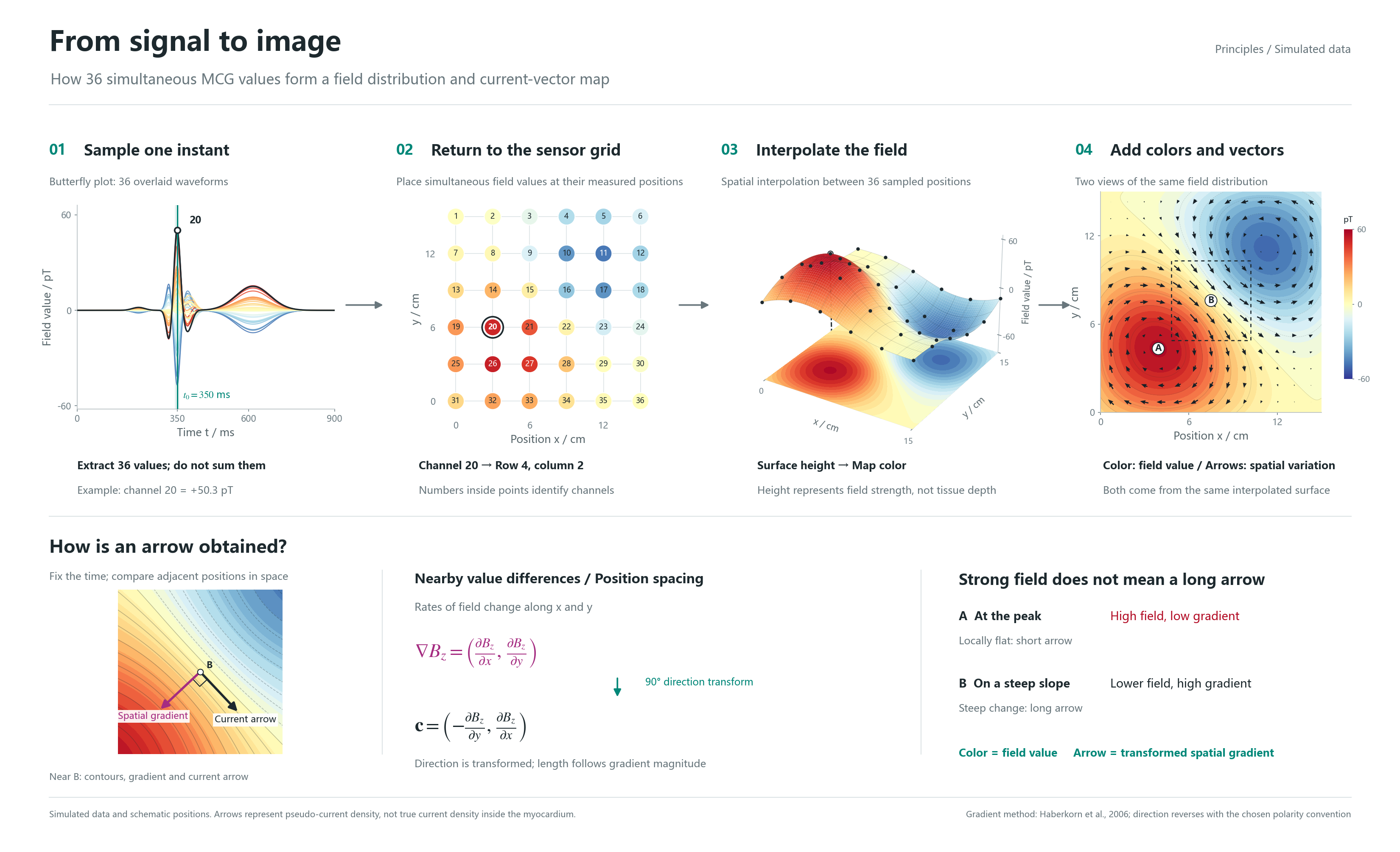
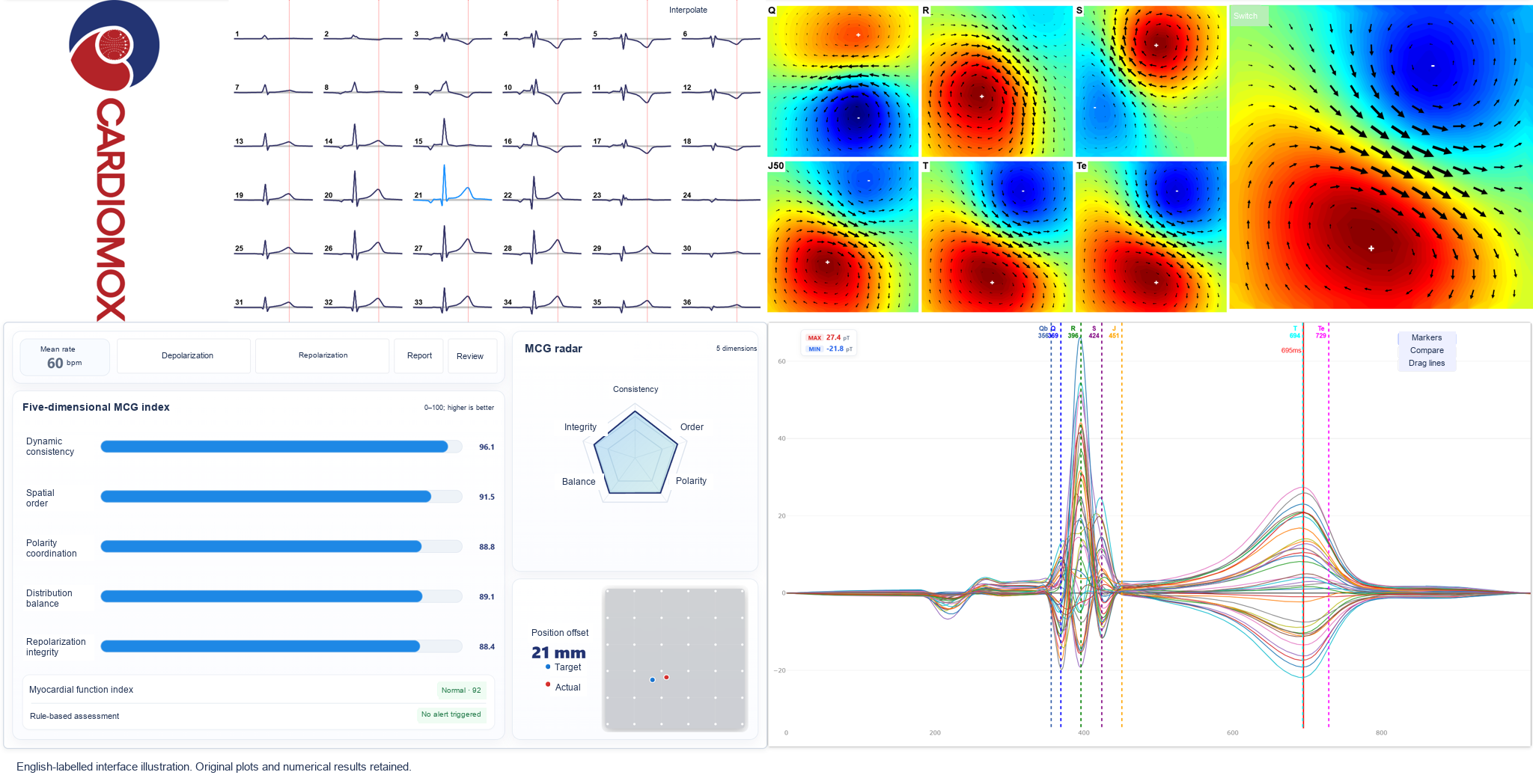
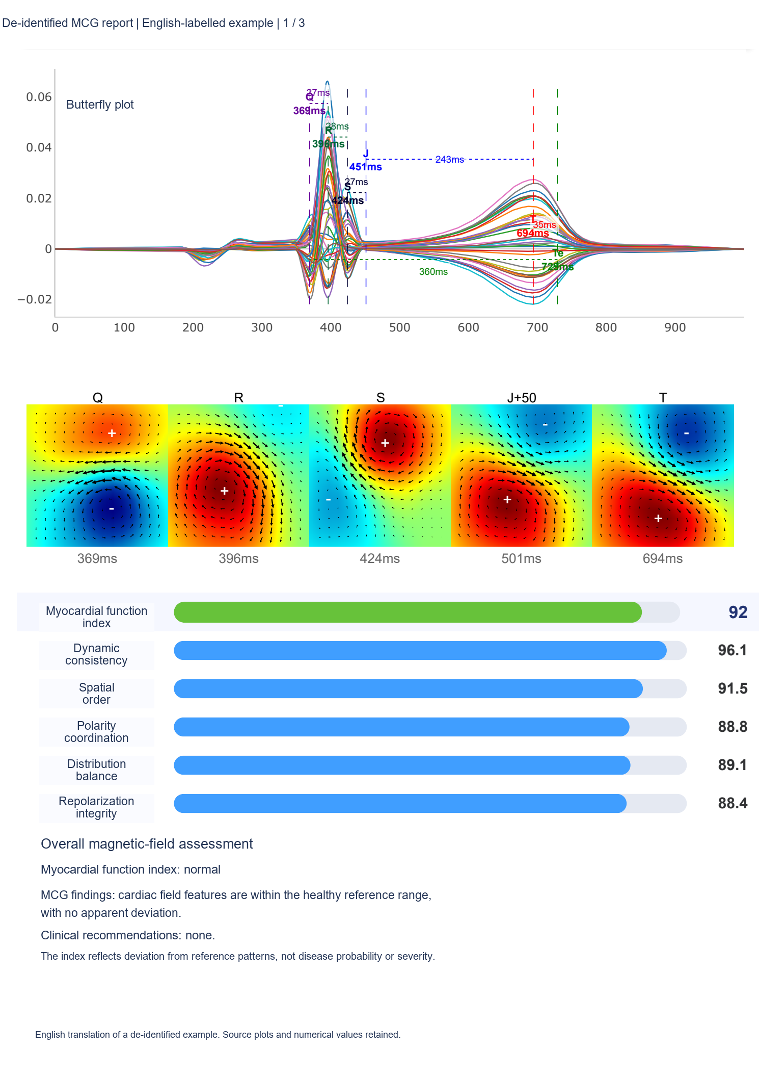
MCG uses magnetic sensors outside the body to detect the weak field generated by cardiac electrical activity. Measurements at multiple positions show both temporal waveforms and spatial field distributions, providing another view of cardiac activity.[3]
Potential and magnetic-field measurements respond differently to internal current sources. They therefore provide partly different, potentially complementary information. MCG is not simply a matter of more measurement points: it measures magnetic information rather than electrical potential.[4]
These are references on measurement principles. The cited FDA document concerns another manufacturer's device, not registration or certification of this product.
Who might discuss MCG with their doctor?
MCG has been studied as an adjunct in the assessment of suspected myocardial ischemia. A confirmed heart-disease diagnosis is not a prerequisite for considering further assessment. A doctor should decide whether MCG belongs in the investigation based on symptoms, risk factors and previous findings.[5]
The following scenarios are for clinical discussion and research context, not a list of approved populations for this product. Registration of MCG-36OPM-01 is pending. Its final intended use will be determined by the approved registration documentation and instructions for use.
- Unexplained occasional or recurrent chest tightness or pain Particularly when symptoms recur, routine tests such as ECG have not explained them, and myocardial ischemia remains a concern.
- Younger adults with chest discomfort alongside irregular schedules or high stress This may include people with frequent late nights, sedentary routines or prolonged stress who also experience chest tightness or pain. Symptoms should not be dismissed because of age or automatically attributed to lifestyle. These circumstances alone are not indications for MCG.
- Chest discomfort during exercise or physical activity Chest tightness or pain while running, exercising or climbing stairs, or a marked decline in exercise tolerance, warrants further evaluation. When ischemia is suspected, the potential adjunctive value of MCG can be discussed with a doctor.[6]
- Smoking history with chest tightness, pain or other symptoms Tobacco exposure is a cardiovascular risk factor. Symptoms warrant assessment for possible ischemia, but smoking history alone does not establish coronary microvascular dysfunction.[7]
- High or borderline blood pressure, glucose or lipids with chest discomfort People with abnormal or borderline values and chest tightness, pain or other clues to ischemia can discuss investigation in the context of their overall health. Abnormal values alone do not establish cardiac damage.
- Angina without significant coronary obstruction, with suspected microvascular dysfunction MCG has been studied in this setting and may be discussed as an adjunctive assessment. The diagnostic pathway requires further validation; MCG cannot establish the diagnosis by itself.[8]
- Known coronary narrowing requiring further assessment for ischemia A doctor may consider the complementary value of MCG when evaluating whether vascular disease is accompanied by ischemia-related functional changes. Studies using other devices do not directly validate this product's diagnostic performance.[9]
- Ischemia assessment when some conventional tests are unsuitable For example, when exercise stress testing is difficult or certain drugs or contrast agents are unsuitable, a doctor should first assess feasible options, then consider whether resting MCG offers adjunctive value. It is not a universal substitute for other tests, nor does this establish suitability of this product for every such patient.
Unsure about your blood pressure, glucose or lipids? Chest discomfort can be assessed as described above. If the only issue is lack of a recent health check, measure blood pressure, glucose and lipids to understand baseline risk; that alone does not establish a need for MCG.[10]
Interpretation: MCG findings must be interpreted in clinical context. MCG alone cannot diagnose or exclude coronary artery disease, and a normal result does not establish that vigorous exercise is safe. The research and health information above do not constitute validation of this product.
Urgent symptoms: Seek emergency medical care for sudden, persistent or worsening chest pain, especially with shortness of breath or cold sweats. Do not wait for an MCG examination.[11]
How should I prepare?
Items containing magnetizable materials such as iron or nickel need attention before the examination. Follow staff instructions to remove items that can be taken off safely, or change clothing containing metal parts, such as metal trouser zippers or bra fasteners, to reduce measurement interference.
Tell staff about metal dentures, intrauterine devices and other implants in advance. This includes older metal dentures or intrauterine devices and any other fixed or implanted metal. The effect on the examination depends on the material and equipment requirements, not simply an item's appearance or age.
Follow staff instructions for removable metal dentures. Do not remove fixed dental work or implanted devices yourself for this examination. A doctor should assess suitability and arrangements. Follow the product instructions and the healthcare institution's requirements.
How long does an examination take with each system?
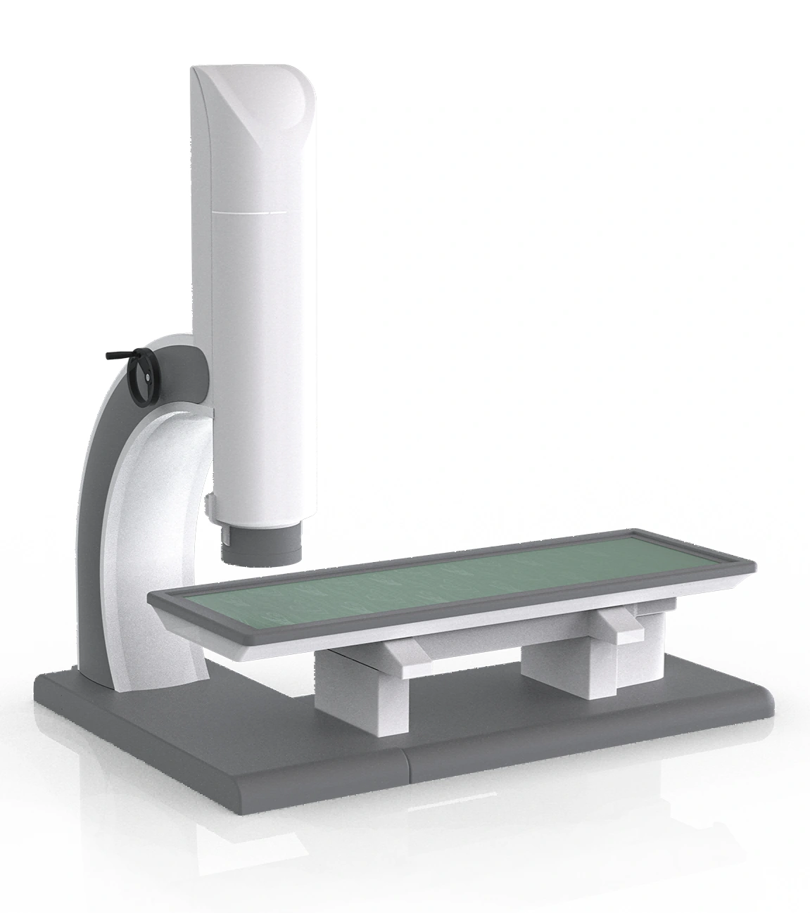
With MCG-36OPM-01, the usual examination takes no more than 3 minutes from lying down on the examination bed to getting off it. This refers to time on the equipment, excluding waiting, changing clothes and clinical interpretation of the report.
Continuous magnetic-signal recording takes approximately 1 minute. This is not the duration of the whole examination. The workflow also includes lying down, initial positioning, entering the shield, automatic fine positioning and withdrawal. Video timers are accelerated; actual time depends on conditions.
The approximately 3-minute workflow applies only to MCG-36OPM-01. MCG 9 SQUID records four regions sequentially, about 1 minute each, with a total examination time of approximately 10–15 minutes depending on local interference. See the MCG 9 workflow.